What’s your diagnosis?
Tucker, a 16 year old Tennesee Walker gelding, presented to the SVEC surgical service with off and on lameness in his right front over the past two years.
What is your diagnosis and next steps/treatments?

The Saginaw Valley Equine Answer
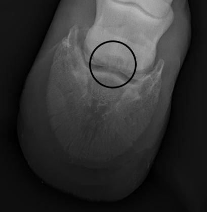
Upon physical examination, Tucker was sensitive to deep palpation of the right front suspensory origin. During the lameness exam, he tested positive to hoof testers in both front feet and was a 3/5 lame at the trot. An abaxial (foot) block was done and he was sound. The radiograph above shows a large navicular cystic lesion in his right front foot.
Over the course of the next year Tucker had a variety of different treatments. The first step was to address shoeing changes and those were successful, but didn’t alleviate all of his symptoms. The next step was coffin joint injections with High Grade HA and corticosteroids. The initial injections kept him sound for about seven months. At this point he came back in for another round of injections. However, this time the benefits didn’t last quite as long.
The decision was made to explore the options of a right front navicular bursoscopy and possible right front neurectomy. For more information on navicular bursoscopy click here. During the bursoscopy, Dr. Brad discussed his prognosis with our surgeon and his owner and they decided to only perform the bursoscopy.
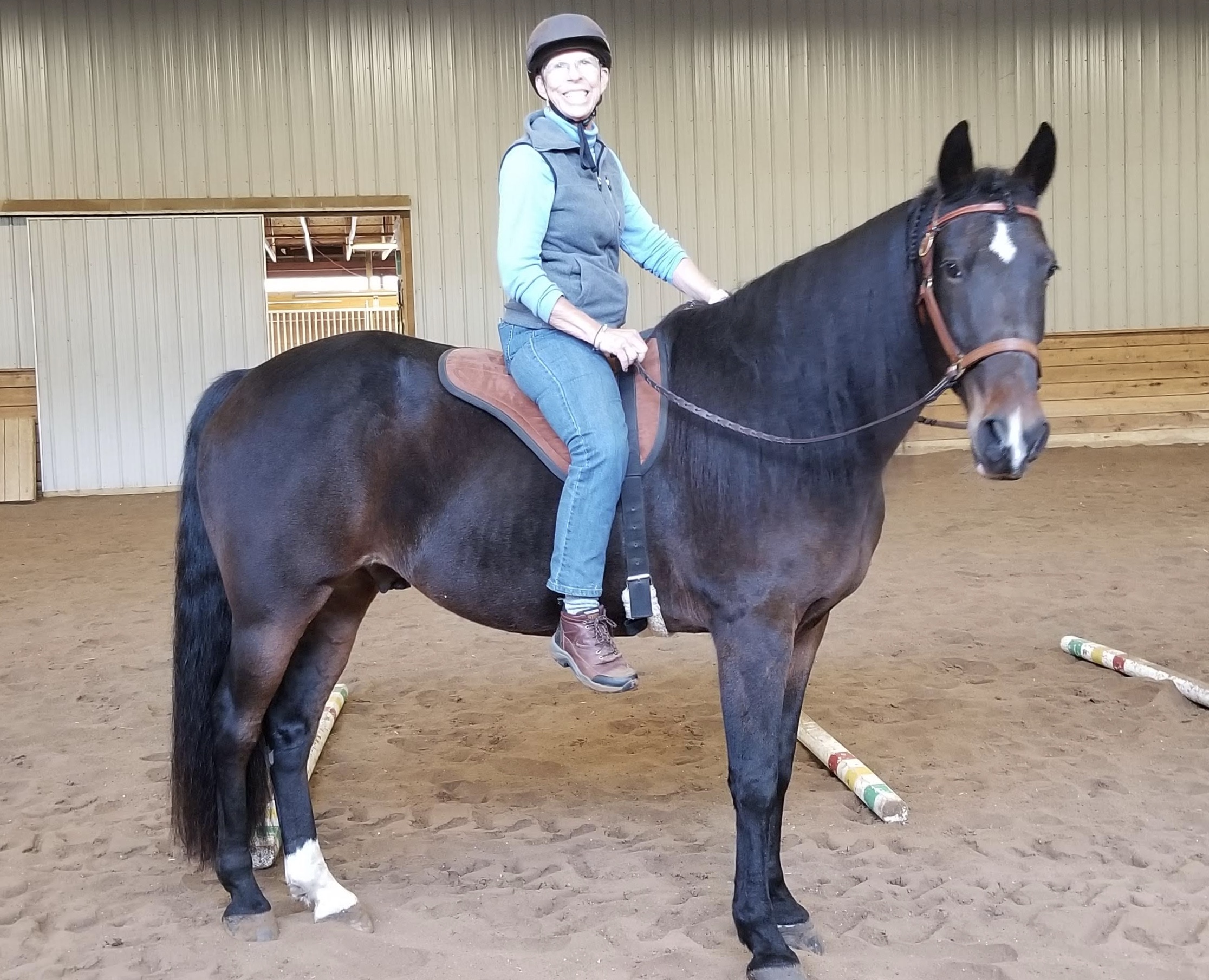
We are happy to report Tucker has recovered well and is doing great!

“The investment in resources both human and financial were worth it. The time I spent soaking, brushing and just hanging with my horse is time I hold dear. We solved many of the worlds problems on our walks. You told me to patient and you were right. The results are better than I hope for. Thank you so much for your care of my friend.”

