What’s your diagnosis?
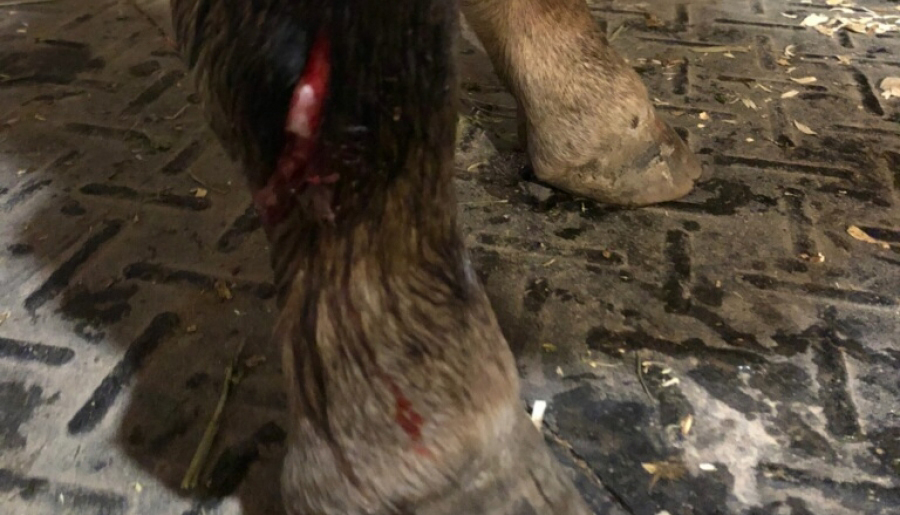
A 2 month old filly was referred to the SVEC surgical service by Dr. Cassandra Shelden with a laceration near her fetlock region on the right hind leg. The day before, she was turned out and somehow got her leg caught up in a wheelbarrow. The owners were present for the accident and immediately cleaned the wound and wrapped the leg with bandage material for the night.
What is your diagnosis and next steps/treatments?


The Saginaw Valley Equine Answer
Diagnostics: Physical Exam – RH fetlock laceration, all other findings within normal limits.
Radiographs and Ultrasound – Within normal limits.
Arthrocentesis (Joint Tap) – a needle was placed in the RH fetlock joint opposite the laceration and 30 ml of sterile saline was then used to pressurize the joint at which time synovial fluid and saline began to leak from the laceration site. The laceration was determined to communicate with the RH fetlock joint.

Sterile fluid being infused with the arthroscope into the joint and expelled from the laceration site. Note how visible the laceration is once the hair is clipped.
Diagnostics: Physical Exam – RH fetlock laceration, all other findings within normal limits.
Radiographs and Ultrasound – Within normal limits.
Arthrocentesis (Joint Tap) – a needle was placed in the RH fetlock joint opposite the laceration and 30 ml of sterile saline was then used to pressurize the joint at which time synovial fluid and saline began to leak from the laceration site. The laceration was determined to communicate with the RH fetlock joint.

Arthroscopic View of the RH Fetlock Joint