What’s your diagnosis?
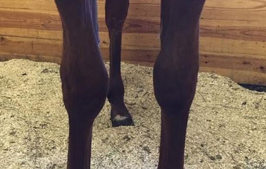
A yearling Paint filly presented to the SVEC surgical service for a lameness and conformation evaluation. Radiographs are pictured below.
What is your diagnosis and next steps/treatments?


The Saginaw Valley Equine Answer
Answer: The filly was diagnosed with carpal valgus in both front limbs.
Diagnostics: Radiographs confirmed that the right front growth plate was longer on the medial aspect (inside) than the lateral aspect (outside) of the knee, causing a carpal valgus (knock kneed) angular limb deformity. A complete ulna was present in both forelimbs, which is an atavism (evolutionary throwback).
Treatment: The filly was placed under general anesthesia for transphyseal screw placement and resection of a portion of the ulna. Intra-operative radiographs guided correct placement of a cortical screw across the growth plate on the medial aspect (inside) of the right forelimb. On the lateral aspect (outside) of the limb, the complete ulna was isolated from surrounding tissues and a 2-3cm section was removed, including the ulnar physis (growth plate). The subcutaneous and skin incisions were sutured and a bandage was placed for recovery.


Update: Two months post surgery
Case #1107 is doing great! A radiograph was taken of the RF carpus (knee) before the screw was removed and revealed no evidence of bone growth over the screw head, allowing standing removal of the screw.
Gallery


