What’s your diagnosis?
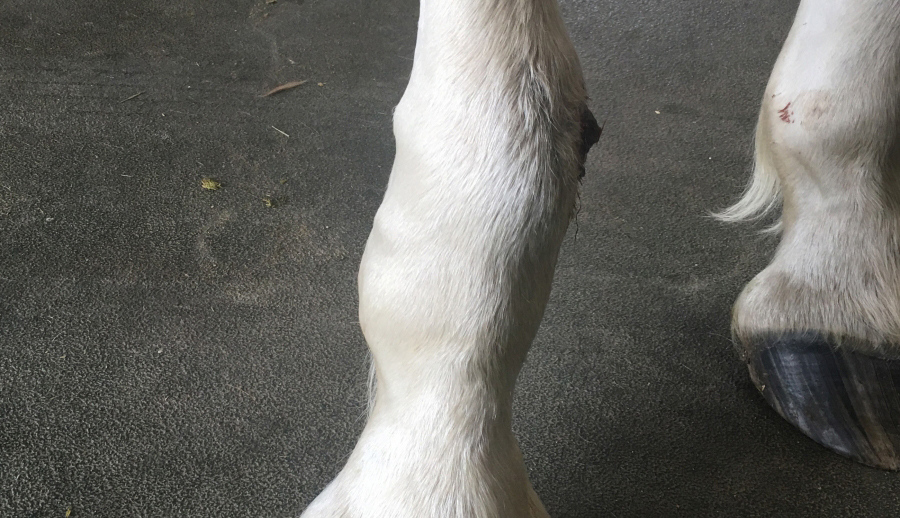
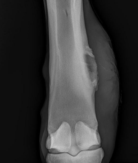
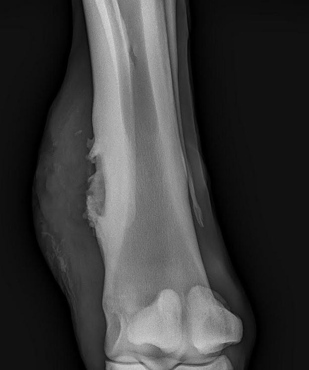
A yearling Belgian filly had a small puncture wound on the right forelimb a few months ago. Now it looks like this.
What is your diagnosis and next steps/treatments?
The Saginaw Valley Equine Answer
Answer: The filly has a sequestrum. A sequestrum is a piece of dead bone tissue occurring within a diseased or injured bone.
Diagnostics: Radiographs of the RF cannon bone revealed significant proliferative bone on the medial aspect of the mid to distal cannon bone. These signs were suspicious for a sequestrum (piece of dead bone) or a foreign body, although neither of these could be distinctly visualized radiographically.
Ultrasound examination revealed a sequestrum (dead bone), but no obvious fluid pockets. There was approximately 3cm of tissue between the skin and the cannon bone.
The area was clipped and cleaned the night before surgery and a bandage was placed to protect the area and decrease swelling prior to surgery.
Pre-operative complete blood cell count revealed a mildly elevated white blood cell count (14K/uL), consistent with infection attributed to the RF wound.
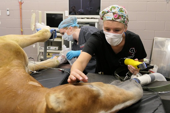
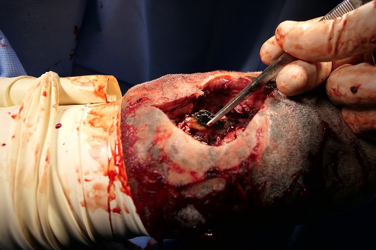
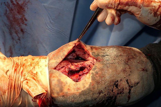
Treatment: The filly was placed under general anesthesia for sequestrum removal from the RF metacarpus. The open wound was removed as a whole section with an elliptical incision. During dissection, a pocket of purulent material opened up and a sample was collected for bacterial culture. A 2-2.5cm section of dead bone (sequestrum) was removed from within the reactive bone. The adjacent bone was curetted to remove infected tissue, but the proliferative bone of the involucrum (bed for the sequestrum) was not removed. Significant amounts of granulation tissue (scar tissue) had to be removed adjacent to the sequestrum in order to allow the skin edges to be apposed. A tourniquet was required intermittently to control bleeding from the vascular granulation tissue. A penrose drain was placed through an exit stab incision distal to the incision. The skin incisions were sutured using stents and a distal limb bandage was placed. Peri-operative antibiotics (ceftiofur, gentamicin) and anti-inflammatory (phenylbutazone) therapy were administered as well as a tetanus toxoid vaccination. Due to discomfort on the RF limb after recovery, additional pain medication (butorphanol) was administered.The following day, a regional limb perfusion with antibiotics (amikacin) was performed and the bandage was changed. There was increased swelling of the incision site, but no concerns. The first dose of long acting antibiotics (Excede) was administered intramuscularly prior to discharge from the hospital.
